

Retinal Imaging 101: Fundus Photos vs. OCT vs. OCT-A (When to Use What)
Modern optometry relies on advanced retinal imaging to detect, diagnose, and manage eye disease earlier and more precisely than ever before. At the New England College of Optometry (NECO), students are trained to use and interpret multiple imaging modalities—each with its own strengths.
This guide breaks down the differences between fundus photography, optical coherence tomography (OCT), and OCT angiography (OCT-A)—and, most importantly, when to use each.
Why Retinal Imaging Matters
The retina provides a direct view into both ocular and systemic health. Conditions such as glaucoma, diabetic retinopathy, and macular degeneration can often be detected before symptoms arise.
Choosing the right imaging modality allows clinicians to:
- Detect disease earlier
- Monitor subtle changes over time
- Guide treatment decisions
- Educate patients with visual evidence
Fundus Photography: The Big Picture
What It Is
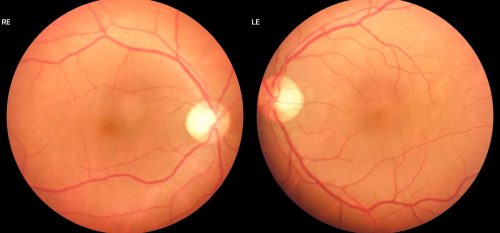
Fundus photography captures a 2D color image of the retina, including the optic nerve, macula, blood vessels, and peripheral retina (depending on the camera type).
Best For
- Baseline documentation
- Monitoring diabetic retinopathy
- Identifying retinal hemorrhages, exudates, or nevi
- Patient education and visual comparison over time
Advantages
- Quick and non-invasive
- Widely available
- Easy for patients to understand
Limitations
- No depth information
- Subtle pathology (e.g., early macular edema) may be missed
When to Use Fundus Photos
Use fundus photography when you need a broad overview or documentation of visible retinal findings. It’s often the first step in retinal evaluation.
Optical Coherence Tomography (OCT): The Cross-Section
What It Is
OCT uses light waves to generate high-resolution cross-sectional images of retinal layers—similar to an optical biopsy.
Best For
- Macular edema
- Age-related macular degeneration (AMD)
- Epiretinal membranes (ERM)
- Glaucoma (RNFL and ganglion cell analysis)
Advantages
- Micron-level resolution
- Detects pathology invisible on fundus exam
- Quantitative measurements for monitoring progression
Limitations
- Limited field of view
- Does not directly visualize blood flow
When to Use OCT
Use OCT when you need to evaluate retinal structure in detail, especially in patients with:
- Reduced or distorted vision
- Diabetes
- Suspected macular disease
- Glaucoma or optic nerve concerns
OCT Angiography (OCT-A): The Vascular Map
What It Is
OCT-A is an extension of OCT that visualizes retinal and choroidal blood flow—without dye injection.
Best For
- Detecting neovascularization
- Evaluating macular ischemia
- Monitoring diabetic retinopathy and AMD
- Identifying vascular abnormalities in the retina
Advantages
- Non-invasive alternative to fluorescein angiography
- Layer-by-layer visualization of blood vessels
- Rapid acquisition
Limitations
- Sensitive to motion artifacts
- Smaller field of view than traditional angiography
- May miss leakage (since it detects flow, not dye pooling)
When to Use OCT-A
Use OCT-A when vascular pathology is suspected, particularly in:
- Diabetic eye disease
- Wet AMD
- Retinal vein occlusions
- Unexplained vision loss with suspected ischemia
Fundus Photos vs. OCT vs. OCT-A: A Quick Comparison
|
Feature |
Fundus Photography |
OCT |
OCT-A |
|
Image Type |
2D surface |
Cross-sectional |
Blood flow mapping |
|
Best For |
Documentation |
Structural analysis |
Vascular assessment |
|
Depth Information |
No |
Yes |
Yes (flow-based) |
|
Detects Blood Flow |
No |
No |
Yes |
|
Typical Use |
Baseline, screening |
Diagnosis, monitoring |
Advanced vascular evaluation |
How NECO Prepares Students in Retinal Imaging
At NECO, students don’t just learn the theory—they gain hands-on experience with industry-standard imaging technology in clinical settings. Through direct patient care, students develop the ability to:
- Select the appropriate imaging modality based on symptoms and risk factors
- Interpret imaging results accurately
- Integrate findings into comprehensive care plans
- Communicate results effectively to patients and colleagues
This real-world training ensures NECO graduates are prepared to deliver evidence-based, technology-driven eye care from day one.
Final Takeaways: Choosing the Right Tool
- Start with fundus photography for documentation and overall assessment
- Use OCT when you need detailed structural information
- Turn to OCT-A for vascular insights without invasive testing
Each modality plays a distinct role—and knowing when to use each is a key clinical skill.
Explore Clinical Training at NECO
Interested in mastering the latest diagnostic technologies in optometry? Learn more about NECO’s Doctor of Optometry program and how immersive clinical training prepares students to lead in a rapidly evolving field of eye care.