Ocular Surface Disease 101: Dry Eye Workup with TBUT, MGD Grading & Staining
 Ocular surface disease (OSD), commonly referred to as dry eye disease, is one of the most frequently encountered conditions in clinical optometry. With increasing screen time, aging populations, and environmental factors, dry eye is becoming more prevalent—and more complex to diagnose and manage.
A thorough dry eye workup is essential for identifying underlying causes and guiding effective treatment. Key components include Tear Break-Up Time (TBUT), meibomian gland dysfunction (MGD) grading, and ocular surface staining.
What Is Ocular Surface Disease?
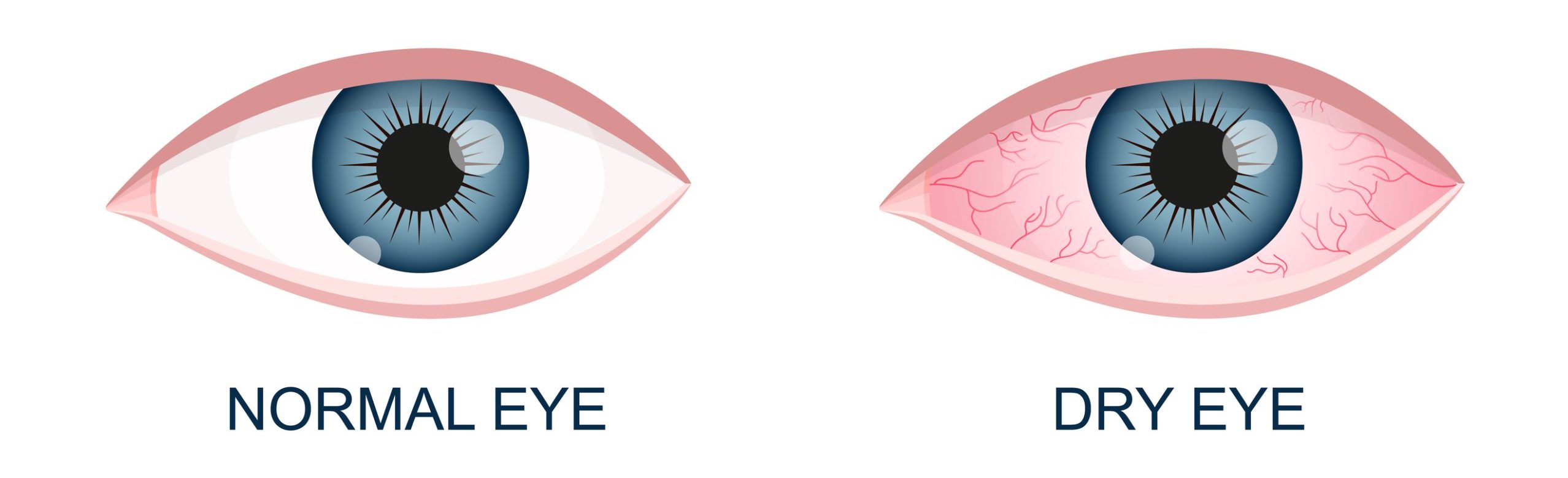
Ocular surface disease is a multifactorial condition characterized by tear film instability, inflammation, and damage to the ocular surface. Patients may experience:
- Dryness or irritation
- Burning or stinging
- Fluctuating or blurry vision
- Foreign body sensation
- Excess tearing (a paradoxical response)
OSD is often divided into two primary categories:
- Aqueous-deficient dry eye
- Evaporative dry eye (commonly associated with MGD)
Understanding the root cause is critical for targeted treatment.
Step 1: Tear Break-Up Time (TBUT)
What Is TBUT?
Tear Break-Up Time (TBUT) measures the stability of the tear film. It is one of the quickest and most informative tests in a dry eye evaluation.
How It Works
After instilling fluorescein dye into the eye, the clinician observes the tear film under cobalt blue light and measures the time between a blink and the first appearance of a dry spot.
Interpreting TBUT
- Normal: ≥10 seconds
- Borderline: 5–9 seconds
- Abnormal: <5 seconds
Short TBUT indicates tear film instability, often associated with evaporative dry eye and meibomian gland dysfunction.
Step 2: Meibomian Gland Dysfunction (MGD) Grading
What Is MGD?
Meibomian glands, located in the eyelids, secrete lipids that prevent tear evaporation. Dysfunction of these glands is a leading cause of dry eye disease.
Clinical Evaluation
MGD assessment typically includes:
- Gland expressibility
- Quality of meibum (clear vs. turbid or toothpaste-like)
- Lid margin appearance (telangiectasia, thickening, irregularity)
MGD Grading Scale (Simplified)
- Grade 0: Clear meibum, normal function
- Grade 1: Cloudy meibum with mild obstruction
- Grade 2: Turbid, thickened secretions with moderate obstruction
- Grade 3: Inspissated glands, little to no secretion
Why It Matters
MGD is responsible for the majority of evaporative dry eye cases. Identifying and grading its severity helps guide treatment options such as:
- Warm compresses
- Lid hygiene
- In-office therapies (e.g., thermal pulsation)
Step 3: Ocular Surface Staining
Purpose of Staining
Ocular surface staining reveals damage to the corneal and conjunctival epithelium, highlighting areas of cell loss or compromise.
Common Dyes Used
- Fluorescein: Highlights corneal defects
- Lissamine green: Stains devitalized conjunctival cells
- Rose bengal: Less commonly used due to irritation
Interpreting Staining Patterns
Different staining patterns can provide diagnostic clues:
- Inferior corneal staining: Often linked to exposure or evaporative dry eye
- Interpalpebral staining: Suggests environmental or evaporative causes
- Diffuse staining: May indicate more severe or advanced disease
Grading scales (e.g., Oxford or NEI) are commonly used to standardize findings.
Putting It All Together: A Comprehensive Dry Eye Workup
A structured approach to dry eye evaluation ensures accurate diagnosis and effective management:
- Patient history and symptom assessment
- TBUT to evaluate tear stability
- MGD grading to assess gland function
- Ocular surface staining to detect tissue damage
Additional tests may include:
- Schirmer testing
- Tear osmolarity
- Meibography
Why Early Detection Matters
Untreated ocular surface disease can significantly impact quality of life and visual performance. It can also affect outcomes for contact lens wear and ocular surgery. Early identification allows clinicians to:
- Prevent disease progression
- Improve patient comfort
- Enhance visual quality
Clinical Training at New England College of Optometry
At New England College of Optometry, students receive comprehensive training in diagnosing and managing ocular surface disease using advanced clinical techniques and technologies. Through hands-on patient care, students develop the skills needed to:
- Perform detailed dry eye evaluations
- Interpret diagnostic findings
- Create personalized treatment plans
Dry eye disease is more than a minor irritation—it is a chronic, multifactorial condition that requires careful evaluation and management. By mastering core diagnostic tools like TBUT, MGD grading, and ocular surface staining, clinicians can provide more precise and effective care.