
OCT-A, Explained: Reading Flow, Artifacts & Pathology
In today’s rapidly advancing world of eye care, OCT angiography (OCT-A) has become one of the most exciting innovations in retinal imaging. This technology allows clinicians—and NECO students—to visualize retinal vasculature and blood flow in remarkable detail, providing crucial insights into diseases that affect vision.
OCT Angiography is an advanced form of Optical Coherence Tomography (OCT), a tool that uses light waves to take high-resolution, cross-sectional images of the retina. What sets OCT-A apart is its ability to capture blood flow without dye injections or invasive procedures.
By comparing sequential OCT scans, OCT-A maps the movement of red blood cells through retinal and choroidal vessels, creating detailed, 3D visualizations of ocular circulation. This enables clinicians to detect subtle changes associated with eye diseases such as:
-
Diabetic Retinopathy (DR)
-
Age-related Macular Degeneration (AMD)
-
Glaucoma
-
Retinal Vein Occlusions
Because it’s noninvasive and highly sensitive, OCT-A provides early diagnostic insight—often before patients experience symptoms—making it one of the most powerful imaging tools in modern optometry.
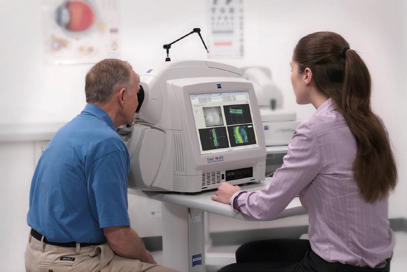
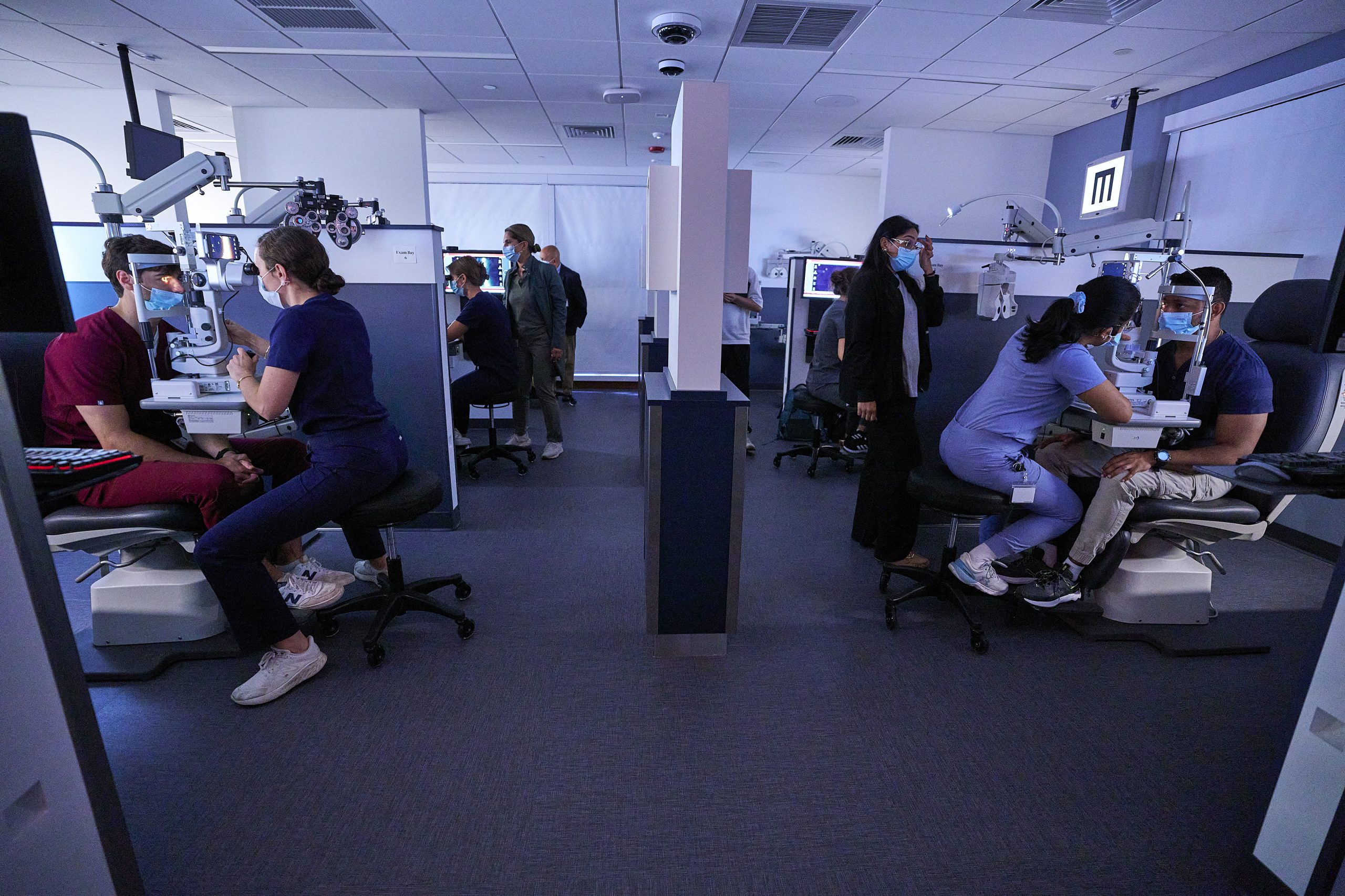
At the New England College of Optometry (NECO), students go beyond theory—they learn OCT-A fundamentals through direct, hands-on training. From mastering OCT-A basics to understanding flow signal interpretation, segmentation layers, and artifacts, NECO prepares future optometrists to confidently apply this technology in clinical settings
Layers, Slabs & What “Flow” Really Means
OCT-A uses high-speed OCT and motion correction to detect the movement of red blood cells, creating en face (top-down, map-like) images through layered “slabs.” Each slab corresponds to a specific vascular network and provides unique diagnostic insights into ocular health.
Key segmentation layers include:
-
Superficial plexus – the inner retinal capillary network surrounding the ganglion cell layer.
-
Deep plexus – located in the inner nuclear layer, representing deeper retinal circulation.
-
Outer retina – normally avascular; any detected flow may indicate macular neovascularization or other pathology.
-
Choriocapillaris – the thin vascular layer beneath the retinal pigment epithelium, crucial for assessing choroidal circulation.
Interpreting these layers requires attention to projection artifacts (false signals projected from superficial layers) and segmentation errors (incorrect layer boundaries). Both can distort visualization and must be corrected for accurate assessment.
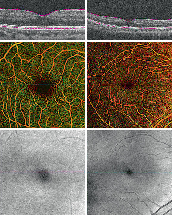
Students also learn to evaluate FAZ size (foveal avascular zone) and patterns of non-perfusion or ischemia clues, which can signal early microvascular compromise in diseases such as DR or macular telangiectasia. Through hands-on imaging and faculty guidance, NECO trainees gain confidence in analyzing OCT-A’s layered data to distinguish normal anatomy from pathology.
Common Artifacts & How to Avoid Misreads
Even expertly acquired OCT-A scans can show artifacts that complicate interpretation. Recognizing and correcting these is a key component of clinical training.
Common artifacts include:
-
Motion artifact – caused by patient movement, leading to duplicated or distorted vessels.
-
Blink lines – dark horizontal gaps from interrupted scans during blinking.
-
Media opacity – cataracts or corneal haze producing masking or signal loss.
-
Decorrelation noise – random speckle or false-positive flow signal due to scan instability.
-
Segmentation errors – misaligned boundaries that place flow data in the wrong retinal layer.
To minimize these issues, NECO students learn to obtain repeat scans, verify segmentation accuracy, and always confirm with structural OCT before forming conclusions. When significant abnormalities such as CNV or macular non-perfusion are suspected, students are trained to apply appropriate referral thresholds to ensure timely specialist evaluation.

By mastering artifact management, interpretation accuracy, and cross-confirmation techniques, future optometrists trained at NECO are well prepared to use OCT angiography confidently in both academic and clinical practice.
Learning OCT-A at NECO
As part of NECO’s state-of-the-art clinical training, students are introduced to OCT and OCT-A technology early in their education. Through both classroom instruction and hands-on clinical experience, they develop the skills needed to:
-
Acquire and interpret OCT-A images with accuracy
-
Identify normal vs. pathological vascular patterns in the retinal vasculature
-
Integrate OCT-A data into comprehensive patient care plans
-
Communicate findings effectively with patients and other healthcare professionals
This structured exposure ensures NECO graduates are confident in using OCT angiography—one of the most powerful, noninvasive imaging tools in modern optometric care—to deliver high-quality, evidence-based patient management from day one of their careers.
Discover more about our clinics, training, and technology at www.neco.edu