Humphrey Visual Field (HVF) Basics: 24-2 vs 10-2, Reliability & Glaucoma Patterns
Visual field testing is a cornerstone of modern optometric care, especially in the diagnosis and management of glaucoma. Among the most widely used tools is the Humphrey Visual Field (HVF) analyzer, which provides detailed, quantitative insight into a patient’s functional vision.
For students, residents, and clinicians alike, understanding HVF basics—including the differences between 24-2 and 10-2 testing strategies, how to assess reliability, and how to recognize glaucomatous patterns—is essential for delivering high-quality patient care.
What Is a Humphrey Visual Field Test?
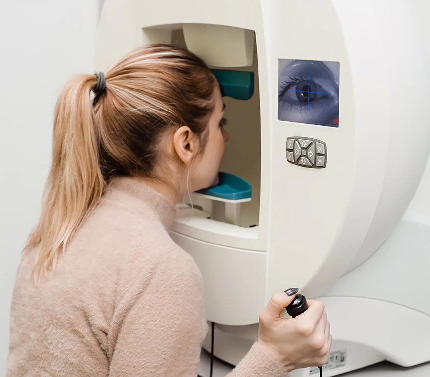
The Humphrey Visual Field (HVF) test is an automated perimetry exam that measures a patient’s central and peripheral vision. It maps light sensitivity across the visual field and detects defects that may not yet be noticeable to the patient.
HVF testing is especially critical for:
- Early detection of glaucoma
- Monitoring disease progression
- Evaluating neurological conditions affecting vision
24-2 vs 10-2: What’s the Difference?
Two of the most commonly used HVF testing protocols are the 24-2 and 10-2. Each serves a distinct clinical purpose.
HVF 24-2
The 24-2 test evaluates a broad area of the central visual field (24 degrees from fixation), using a grid of test points spaced 6 degrees apart.
Best for:
- Initial glaucoma screening
- Monitoring moderate to advanced glaucoma
- Assessing peripheral and mid-peripheral defects
Advantages:
- Wider field coverage
- Efficient for routine clinical use
Limitations:
- Less sensitive to subtle central defects
HVF 10-2
The 10-2 test focuses on the central 10 degrees of vision, with a denser grid (2-degree spacing between points).
Best for:
- Detecting early central glaucomatous damage
- Monitoring macular involvement
- Evaluating patients with suspicious central defects on 24-2
Advantages:
- High resolution in the central field
- Better detection of small or early defects near fixation
Limitations:
- Limited peripheral information
When to Use Each Test
Clinicians often use both strategies together. A common approach:
- Start with 24-2 for general assessment
- Add 10-2 if:
- Central defects are suspected
- The patient has early glaucoma
- Structural imaging (e.g., OCT) suggests macular damage
Increasingly, research shows that glaucoma can affect the central visual field earlier than previously thought—making 10-2 testing more important than ever.
Assessing Test Reliability
Before interpreting any HVF result, it’s critical to evaluate test reliability. Even subtle patient errors can significantly affect results.
Key Reliability Indices
1. Fixation Losses
- Indicates whether the patient maintained steady gaze
- High values suggest poor fixation
2. False Positives
- Occur when the patient responds without a stimulus
- Often described as a “trigger-happy” response pattern
- Can make the field appear better than it is
3. False Negatives
- Occur when the patient fails to respond to a visible stimulus
- May indicate fatigue, inattention, or advanced disease
Acceptable Reliability Guidelines
While not absolute, commonly accepted thresholds include:
- Fixation losses: <20%
- False positives: <15%
- False negatives: <15–20%
Clinical Tip:
Always interpret reliability indices before analyzing the visual field. An unreliable test can lead to misdiagnosis or inappropriate management.
Recognizing Glaucoma Patterns on HVF
Glaucomatous visual field loss follows characteristic patterns due to damage to the retinal nerve fiber layer.
Common HVF Patterns in Glaucoma
1. Nasal Step
- Defect respecting the horizontal midline
- Often an early sign of glaucoma
2. Arcuate Scotoma
- Arc-shaped defect extending from the blind spot
- Follows the nerve fiber layer
3. Paracentral Scotoma
- Small defect near fixation
- May be missed on 24-2 but detected on 10-2
4. Temporal Wedge
- Less common, but may occur in advanced disease
Key Global Indices
In addition to pattern recognition, clinicians rely on summary metrics:
- Mean Deviation (MD): Overall depression of the visual field
- Pattern Standard Deviation (PSD): Irregularities in the field (localized defects)
- Visual Field Index (VFI): Percentage of visual function remaining
These indices help track progression over time and guide treatment decisions.
Why HVF Mastery Matters
For optometry students and clinicians, mastering HVF interpretation is not just an academic exercise—it directly impacts patient outcomes.
At institutions like New England College of Optometry, students gain hands-on experience with advanced diagnostic technologies, preparing them to:
- Detect glaucoma earlier
- Monitor progression more precisely
- Deliver evidence-based care
Understanding the nuances of Humphrey Visual Field testing—from choosing between 24-2 and 10-2 to evaluating reliability and identifying glaucomatous patterns—is essential for modern optometric practice. As technology evolves and our understanding of glaucoma deepens, clinicians must continue refining their skills to ensure the best possible care for their patients.