Corneal Tomography vs. Topography in Ectasia Screening
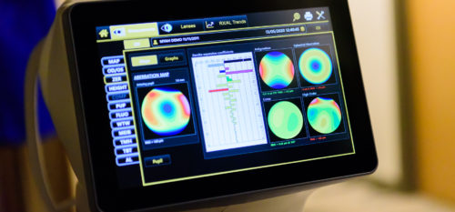
Accurate imaging is central to keratoconus screening and identifying patients at risk for post-LASIK ectasia. Corneal topography has long served as the standard for evaluating surface curvature, but modern corneal tomography powered by Scheimpflug and OCT-based systems offers a deeper and more comprehensive assessment. At the New England College of Optometry (NECO) and within NECO clinics, understanding how each modality contributes to diagnosis and surgical decision-making is essential for patient safety and early disease detection.
What Topography Tells You (and What It Misses)
Corneal topography uses Placido-based reflection to generate a 2D anterior-surface map of the cornea. By analyzing anterior curvature patterns and keratometric indices, clinicians can visualize surface irregularities that support keratoconus screening and refractive surgery planning.
Topography is particularly useful for:
-
Recognizing asymmetric bow-tie patterns
-
Identifying inferior steepening
-
Assessing pre- and post-refractive surgery changes
-
Monitoring surface-level progression
-
Fitting specialty lenses where curvature data and repeatability matter
However, topography is limited by its dependence on a smooth, stable tear film; tear film impact can alter surface readings. It provides no posterior data, a critical limitation: no posterior data. Because it cannot image the posterior corneal surface or map corneal thickness, early ectatic changes may go undetected.
Tomography Advantages for Early Disease
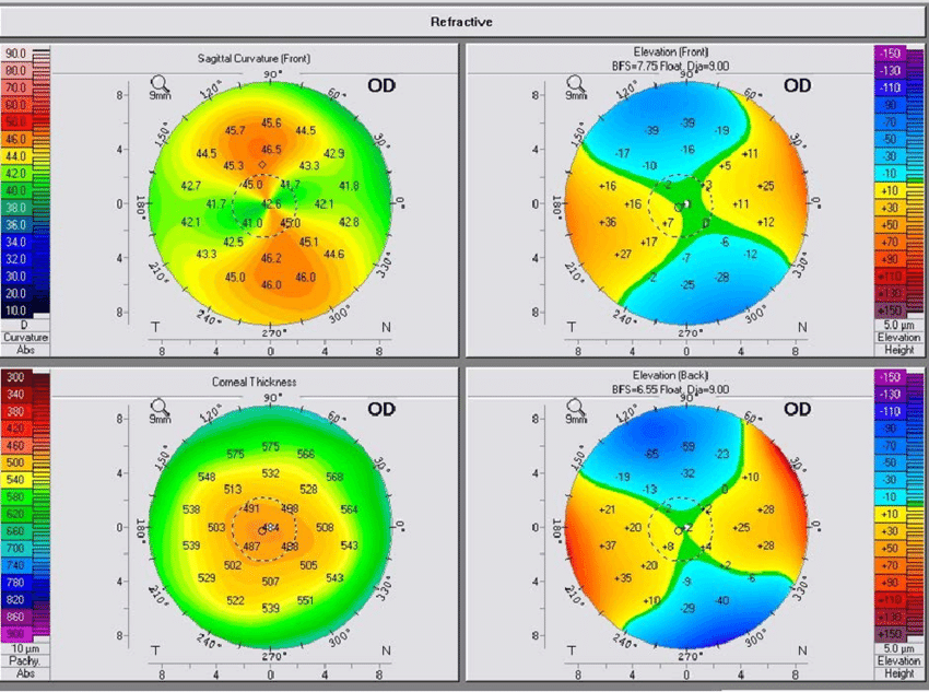
Corneal tomography creates a 3D reconstruction of both the anterior and posterior corneal surfaces and generates pachymetry maps throughout the cornea. Using Scheimpflug or OCT acquisition, tomography captures true corneal shape and volume, including the earliest signs of structural compromise.
Key advantages include:
-
Pachymetric progression and thickness spatial profile to evaluate thinning patterns
-
Posterior elevation and posterior float, allowing detection of changes not visible on topography
-
Enhanced ectasia display and indices such as Belin/Ambrosio and BAD-D for comprehensive risk profiling
-
True net power calculations that incorporate both surfaces
-
Improved identification of surgical candidacy for LASIK/PRK
-
Data-driven guidance for determining follow-up intervals in early or borderline cases
These metrics allow clinicians to detect early keratoconus or post-LASIK ectasia risk before anterior-surface signs appear.
Clinical Applications in Ectasia Screening
Tomography provides a more complete picture of corneal biomechanics and structure, strengthening both screening and long-term monitoring:
-
Early identification of subtle posterior elevation or localized thinning
-
Pre-operative refractive screening to avoid postoperative ectasia
-
Longitudinal monitoring of shape, thickness, and risk indices
-
Integration of pachymetry maps and elevation data for evidence-based risk assessment
At NECO clinics and the NECO Center for Eye Care, students and clinicians learn to interpret both topographic and tomographic data, integrating imaging findings with clinical history, patient symptoms, and long-term management strategies.
Topography vs. Tomography: A Quick Comparison
| Feature | Topography | Tomography |
|---|---|---|
| Data Type | 2D anterior curvature map | 3D reconstruction (anterior + posterior) |
| Imaging Method | Placido reflection | Scheimpflug, OCT, or slit-scanning |
| Corneal Layers | Anterior surface only | Anterior, posterior, and pachymetric |
| Key Metrics | Keratometric indices, bow-tie patterns, surface steepening | Posterior elevation, pachymetric progression, BAD-D, true net power |
| Ectasia Detection | Moderate; limitation: no posterior data | High sensitivity, especially early stage |
| Clinical Use | Contact lens fitting, refractive surgery planning | Comprehensive screening, surgical candidacy, risk assessment, progression monitoring |
The Future of Corneal Imaging at NECO
As tomography becomes standard in clinical practice, NECO continues expanding hands-on training and research opportunities to prepare future clinicians for advanced disease detection. Students gain experience interpreting complex datasets such as anterior curvature, pachymetry maps, posterior elevation, and BAD-D metrics and translating them into practical patient-care decisions within NECO clinics.
A faculty member in anterior segment disease explains that understanding the difference between surface appearance and deeper structural change is foundational for preventing vision loss. Tomography reveals those deeper changes earlier, sometimes years before symptoms develop.
Takeaway
Topography and tomography both play essential roles in modern corneal care. Topography remains indispensable for analyzing anterior curvature and supporting contact lens fitting, but tomography’s 3D imaging provides the most sensitive and complete evaluation for keratoconus screening, post-LASIK ectasia risk, and early structural change.
Learn more about clinical training and imaging technology at NECO by visiting the NECO clinical website: https://necoeyecare.org.